What causes cartilage lesions ?
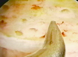
Cartilage lesions can be caused by trauma (accident), microtrauma (overload), or degeneration (age and wear). The pain in such an injury is triggered by the defect in the cartilage and the excessive pressure on the underlying bone, leading to bone marrow swelling and pain. The associated swelling can also contribute to joint pain.
Can we treat all cartilage lesions ?
Traumatic and micro-traumatic injuries are quite treatable, but it is more challenging for degenerative injuries. Patients over the age of 50 are less suitable candidates for a biological option. If cartilage injuries are left untreated, the cartilage layer can gradually erode, leading to osteoarthritis and progressive loss of mobility.
Treatment options
Currently, there are various therapies available depending on age, severity, and activity level.
- Medication: Classic pain relief (paracetamol and anti-inflammatory drugs) and glucosamine preparations are often used in the initial phase of treatment or after surgery.
- Weight reduction: Reducing the load on the knee joint can lead to a significant decrease in pain. If you are significantly overweight (BMI over 35), a referral to the obesity clinic is typically recommended.
- Adapted Activities and Physiotherapy: Activities like cycling, swimming, and low-impact exercises such as fitness are excellent options for the knee joint
Injections with cortisone, hyaluronic acid, plateletrich plasma (PRP) and microfragmented fat
Osteoarthritis, a common issue, arises from various causes including sports injuries, abnormal knee shape, genetic factors, excessive use, and obesity. Treatment focuses on strengthening the knee, reducing risk factors, and managing inflammation.
- Physiotherapy and supportive measures, like weight reduction and basic pain relief, are effective.
- Cortisone injections reduce inflammation and pain, but frequent use may have negative effects on healthy tissues.
- Hyaluronic acid, a natural lubricant, nourishes and protects cartilage. Recent data suggest high-dose injections are as effective as multiple smaller ones. It’s often combined with an antioxidant.
- Platelet-rich plasma (PRP) injections, derived from blood, contain growth factors that promote healing. Typically, three injections are required for an optimal response.
- Fat and stem cell injections are gaining attention, but scientific data is limited. These cells, abundant in abdominal fat, can be harvested and injected to suppress inflammation and aid in repair.
- All injections aim to strengthen and alleviate pain in a worn-out joint, with each having its pros and cons. Common side effects for all injections include pain and swelling at the injection site.
Shaving and Debridement
During keyhole surgery, the loose pieces of cartilage are removed using a shaver. The edges of the cartilage injury are then stabilized.
Microfracture or Ice-Picking
 Microfracture, also known as ice-picking, involves creating small holes in the underlying bone during exploratory surgery. When using the ice-picking technique, the bone marrow seeps through the holes in the injury, forming a blood clot containing stem cells to fill the cartilage injury. These stem cells can then generate repair tissue. This technique is suitable for isolated cartilage injuries up to 1.5 square cm. A recent study has demonstrated that incorporating specific gels and scaffolds can enhance healing after microfracture.
Mosaicplasty
Cartilage and bone plugs are transplanted from another part of the knee to the injury. The diameter and number of plugs may vary depending on the size of the lesion. It is recommended not to use more than 3-4 plugs, which is why this technique is ideal for smaller cartilage injuries where the underlying bone is also affected or injured.
Autologous Chondrocyte Transplantation

In the first step, pieces of cartilage are extracted during keyhole surgery (biopsy). These cartilage pieces are sent to the lab where the cells are cultured for about 10 weeks. In the second step, these cells are then implanted during an open procedure through a small incision. The cells are initially placed onto a collagen membrane that is sutured or adhered into the lesion. This technique is suitable for isolated cartilage lesions ranging from 2 to 10 square cm. Unfortunately, this technique has not been available in Europe since June 2016!
Cartilage Scaffolds
 In cases where both cartilage and bone are affected, an artificial cartilage membrane or scaffold can be implanted. This scaffold enables the restoration of both bone and cartilage in a single operation. Scaffolds are available in the form of plugs or foam.
Metal Implants
 In cases of complex cartilage injuries or in elderly patients, a non-biological solution can be considered. The cartilage damage is covered by a custom-made metal implant for the patient. This form of treatment is currently only available for selected cases.
Rehabilitation
Rehabilitation also plays a very important role in recovery and starts the day after surgery. It is crucial to be well-informed and guided throughout the process. A full rehabilitation period can take up to a year. Sports activities are restricted during the first year and for up to 18 months after the procedure. Additional precautions include wearing a brace for three months and initially refraining from using a brace for two to four weeks.

